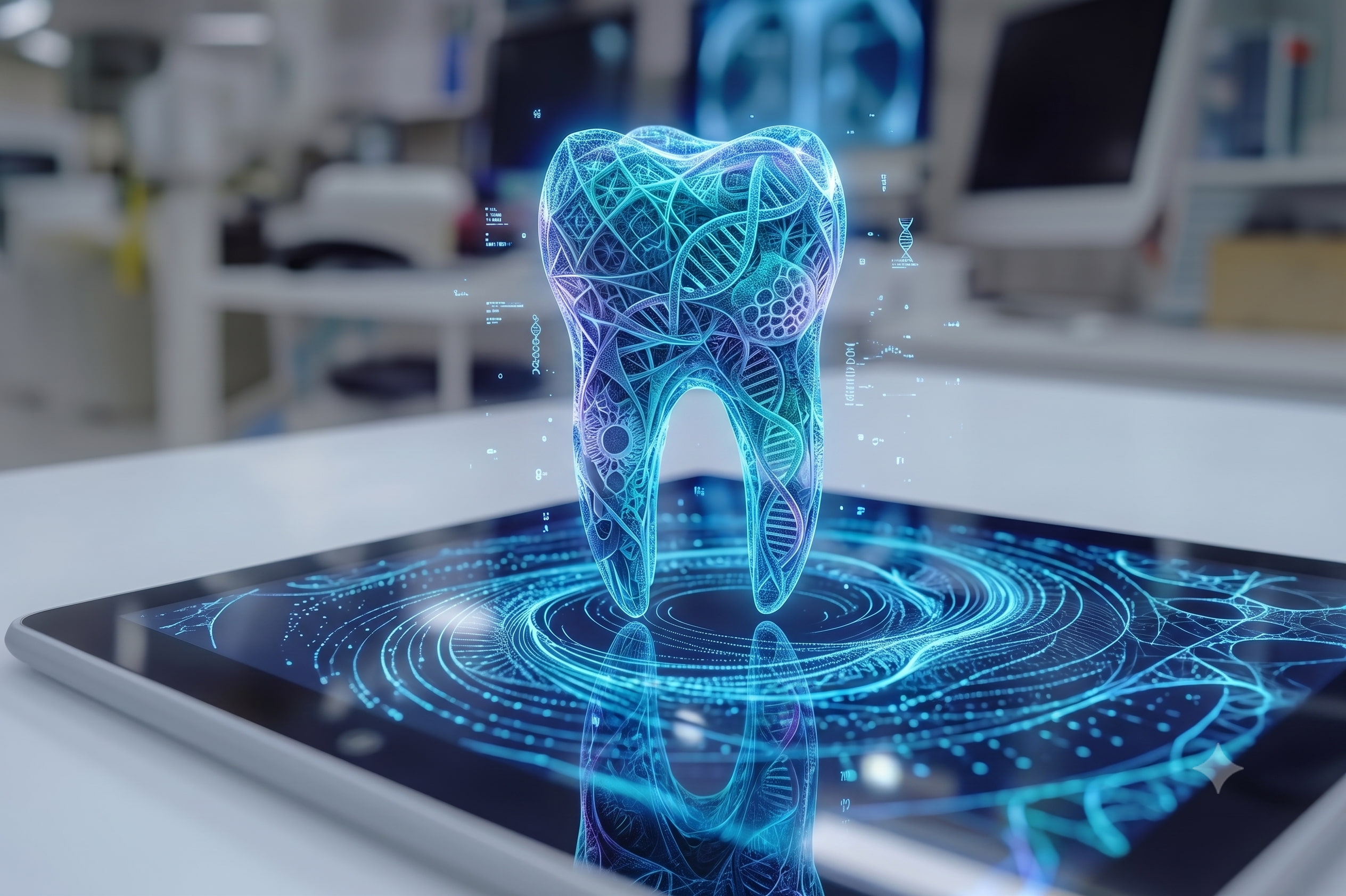
Digital dentistry in Vietnam can make treatment planning more accurate, transparent and easier to explain. It does this by giving the dentist better information before treatment begins—not by guaranteeing how the final result will look or how the body will heal.
For international patients, this distinction matters. A clinic may advertise CBCT imaging, 3D scanning, Digital Smile Design or computer-guided surgery, but a list of equipment says little about how those tools affect an individual treatment plan.
The real value of a digital workflow lies in four questions: what information was captured, who interpreted it, how it changed the treatment decision and how the plan will be checked during treatment.
Quick Answer
Digital dentistry in Vietnam combines three-dimensional imaging, surface scanning and design software to support diagnosis, implant positioning and restoration planning. CBCT, intraoral scanning and Digital Smile Design provide different information. They can reduce avoidable uncertainty, but the outcome still depends on diagnosis, clinical execution, laboratory work, healing and aftercare.
Digital Dentistry Improves Predictability, It Does Not Guarantee an Outcome
Prediction and predictability are not the same thing.
A digital system may allow a dentist to measure available bone, examine the position of anatomical structures, record the bite or simulate a proposed smile. This replaces some visual estimates with measurable information and helps the clinical team identify limitations earlier.
The technology cannot determine every biological or practical outcome in advance. Gum response, bone healing, implant stability, bite forces, patient hygiene and the accuracy of each clinical and laboratory stage can all affect the result.
Digital workflows also contain several opportunities for small errors. A scan must be captured correctly, different datasets may need to be aligned, the plan must be transferred to the mouth and the restoration must still be fitted and adjusted clinically. A computer-generated plan is therefore a guide for treatment, not an automated decision.
This is particularly relevant to computer-guided implant placement. The software may help the dentist plan implant position and design a surgical guide, but the implant placed in the mouth may not correspond perfectly with the virtual position. The dentist still needs to allow appropriate safety margins and respond to the anatomy and implant stability found during surgery.
Modern equipment is most useful when it supports sound clinical judgement. It cannot compensate for an incomplete diagnosis or an unsuitable treatment plan.
What CBCT, Intraoral Scanning and Digital Smile Design Actually Do
The term “3D dental scan” is sometimes used for several different technologies. A CBCT scan and an intraoral scan are not interchangeable, and Digital Smile Design serves another purpose again.
CBCT Shows the Anatomy Beneath the Surface
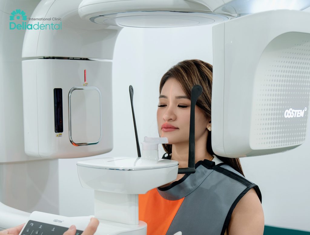
Cone beam computed tomography produces a three-dimensional dataset of the selected dental and maxillofacial area. Unlike a dental photograph or surface scan, CBCT can provide information about the jawbone and nearby anatomical structures.
For implant treatment, the dentist may use it to assess the height and width of available bone, the position of the mandibular canal, the relationship between the upper jaw and the maxillary sinus, and the condition of a proposed implant site. These findings may influence implant length, diameter, angle, surgical approach or whether bone management needs to be considered.
A CBCT scan does not automatically confirm that grafting is required or that an implant will integrate successfully. Its findings must be interpreted with the patient’s examination, medical history, gum condition, bite and restorative needs.
It should also be used when clinically justified. The US Food and Drug Administration’s CBCT guidance notes that CBCT provides three-dimensional information but normally involves more radiation than conventional dental X-rays. It should therefore answer a relevant clinical question rather than be performed simply because a clinic owns the equipment.
An Intraoral Scanner Records Teeth, Visible Gums and Bite Surfaces
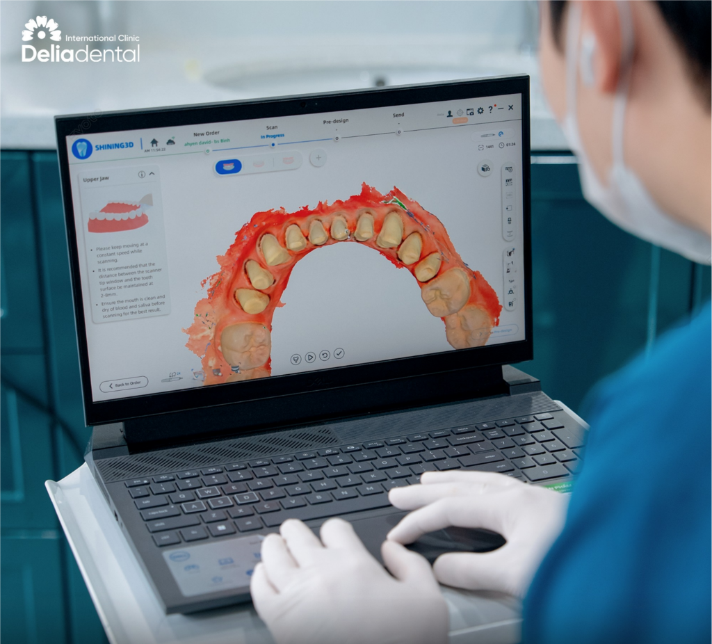
An intraoral scanner captures the visible surfaces inside the mouth and converts them into a digital model. It can record tooth shape, prepared teeth, accessible gum contours, implant scan bodies and the relationship between the upper and lower teeth.
This information may be used to design crowns, veneers, bridges or implant restorations and to transfer measurements to a dental laboratory without relying entirely on a conventional impression.
An intraoral scanner does not see inside the jawbone. It cannot replace CBCT when the dentist needs three-dimensional information about bone or underlying anatomy.
Accuracy also depends on the situation. A short scan of a single tooth is different from recording an entire arch with several implants. Scanner type, operator technique, scan path, moisture, arch length and the position of implant scan bodies can all affect the dataset.
A 2024 systematic review of full-arch intraoral scans found that the evidence remains heterogeneous and that accuracy can vary with the scanner, scanning method and clinical situation. This does not make digital impressions unreliable. It means a clinic should understand when an intraoral scan is appropriate and when further verification or another recording method is needed.
Digital Smile Design Creates a Preview for Discussion
Digital Smile Design uses photographs, facial references and dental data to help the patient and dentist discuss the intended appearance of a restoration. It may be used to explore tooth length, width, proportion, symmetry and the relationship between the teeth, lips and face.
Its strongest role is communication. A patient considering crowns or veneers can respond to a proposed design before the permanent restorations are completed. The dentist and dental technician also gain a clearer shared reference for the intended result.
The preview is not a contractual image of the final teeth. Software cannot reproduce every change in gum tissue, lip movement, material translucency, lighting, bite or healing. The design may also need to change once the teeth and gums are examined directly.
For this reason, mock-ups, temporary restorations and trial fittings remain important. They allow the appearance, speech, bite and comfort of a design to be assessed in the mouth rather than only on a screen.
How a Digital Workflow Changes Treatment Decisions
Digital tools become more useful when their information is connected rather than reviewed as separate scans.
For Dental Implant Treatment
In a digital implant workflow, CBCT may provide information about the jawbone and anatomical boundaries, while an intraoral scan records the surface of the teeth, gums and bite. Planning software can combine relevant datasets so that the proposed implant position is considered in relation to both the available bone and the intended final tooth.
This is known as restoratively driven planning. Instead of selecting an implant position from the bone image alone, the dentist considers where the final crown or bridge needs to emerge, how it will meet the opposing teeth and whether the patient will be able to clean it.
When appropriate, the plan may be transferred through a surgical guide. However, not every implant case requires guided surgery, and a guide does not remove the need for surgical judgement. The dentist must still assess infection, gum thickness, mouth opening, bone condition and whether adequate primary stability can be achieved.
This is also why a 3D dental scan before implants may change an earlier proposal. As Delia Dental Clinic explains in its guide to why implant quotes can change after CBCT, three-dimensional imaging may reveal limitations that were not visible in photographs or a preliminary two-dimensional X-ray.
For Crowns, Veneers and Smile Rehabilitation
For cosmetic and restorative work, photographs and Digital Smile Design help establish the intended visual direction. The intraoral scan records the teeth and bite, while CAD/CAM software or a dental laboratory uses the information to design the restorations.
The design still needs clinical checkpoints. A temporary restoration may reveal that the proposed teeth feel too bulky, affect speech or interfere with the bite. A trial fitting allows the dentist to check contact points, margins, shape, shade and occlusion before final placement.
Delia Dental Clinic ’s guide to natural-looking porcelain crowns in Vietnam shows how Digital Smile Design, scanning, temporary teeth and trial fitting can form separate stages of one restorative workflow. The scan begins the design process; it does not replace the later checks.
What Can Be Assessed Before You Travel to Vietnam?
Digital records can make a remote consultation more useful, particularly when a patient is comparing treatment in another country.
Recent X-rays, original CBCT DICOM files, clear dental photographs, treatment records and an existing quote may help a clinic understand the likely treatment area and identify possible options. If an intraoral scan has already been taken, ask whether the clinic can open the original file format before sending it.
The team may then discuss a preliminary treatment direction, likely number of stages, possible travel timeline and initial cost range. They may also identify which records are missing or no longer current.
However, a remote assessment has limits. The dentist cannot palpate the gums, test tooth mobility, examine the bite fully or evaluate every clinical sign from digital files. An old CBCT may no longer represent a site that has changed following extraction, infection or recent treatment. A scan may also have the wrong field of view or insufficient image quality.
The final diagnosis and treatment plan should therefore be confirmed after an in-person examination and any clinically necessary updated imaging. A clinic should be able to distinguish clearly between what the submitted records show and what remains uncertain.
How to Evaluate a Clinic’s Digital Workflow
Patients do not need to judge one scanner brand against another. The more useful test is whether the clinic can explain how digital information is converted into a clinical decision.
| What to check | Stronger answer | Weaker answer |
|---|---|---|
| What did the technology change? | Identifies the finding and the treatment decision it affected | Lists equipment without explaining its purpose |
| Are the datasets connected? | Bone, surface and restorative information are compared where relevant | Each scan is viewed as a separate marketing feature |
| Who approves the plan? | A named dentist interprets and confirms it | “The software decides” |
| How is a preview verified? | Mock-up, temporary restoration or trial fitting is used | The screen preview is treated as the guaranteed result |
| How are errors controlled? | Clinical checks occur at important treatment stages | The system is described as “100% accurate” |
| Can records be provided? | Relevant imaging and treatment details can be handed over | The patient receives little documentation |
A clinic does not need to use every available technology in every case. In fact, recommending a scan without a clear clinical reason is not necessarily a sign of better care.
A stronger answer explains why a particular record is needed, what it can reveal and how its limitations will be managed. The technology should make the treatment rationale easier to understand—not hide it behind technical language.
How Delia Dental Clinic Uses Digital Records in Treatment Planning
Delia Dental Clinic describes the use of CBCT imaging in implant assessment and intraoral scanning, Digital Smile Design and CAD/CAM processes in restorative treatment. These technologies serve different stages rather than functioning as one machine that predicts a result.
Before travelling, international patients can send recent X-rays, existing CBCT files, dental photographs, treatment records or another clinic’s proposal for review. Delia Dental Clinic’s remote implant assessment guide explains that the usefulness of these records depends on their type, date, quality and coverage.
The initial review can help identify a likely treatment direction, possible timeline, cost components and information still required. It should not be treated as final confirmation of implant position, immediate-loading suitability, grafting, restoration design or complete price.
After the patient arrives at Delia Dental Clinic’s Hanoi or Ho Chi Minh City clinic, the dentist examines the teeth, gums, bite and medical history. Existing imaging is reviewed, and new imaging or scanning is performed when clinically required. The final plan is then confirmed before treatment proceeds.
For crown, veneer and larger restorative cases, the digital design is followed by physical checkpoints. Temporary restorations and trial fittings give the patient and clinical team an opportunity to assess appearance, bite, speech and comfort before the permanent restoration is completed.
Send your recent X-rays or CBCT files, clear dental photos and any existing treatment plan via WhatsApp or Messenger. Delia Dental Clinic ’s team in Hanoi or Ho Chi Minh City can review what the records show, identify missing information and explain which parts of the plan still require an in-person examination.
Frequently Asked Questions
What technology is used for dental implants?
Dental implant planning technology may include dental X-rays, CBCT, implant-planning software and an intraoral scanner. A surgical guide may be designed when clinically appropriate. Not every patient needs every tool, and the treating dentist should explain how each one affects the proposed implant treatment.
Is a 3D dental scan the same as a CBCT scan?
No. An intraoral scanner records visible surfaces such as teeth, accessible gums and bite relationships. CBCT uses X-ray imaging to produce three-dimensional information about the jawbone and surrounding anatomy. The two datasets may complement each other, but one does not automatically replace the other.
Can Digital Smile Design show exactly what my final teeth will look like?
No. Digital Smile Design provides a visual planning and communication reference. The final appearance may be influenced by the condition of the teeth and gums, material selection, laboratory work, clinical adjustments and healing. Mock-ups and trial fittings provide additional opportunities to evaluate the design.
Does computer-guided implant surgery remove all risk?
No. Guided surgery can help transfer a virtual plan to the mouth, but it does not eliminate positional deviation, surgical complications or biological risks. Accurate records, suitable planning, clinician experience, safety margins and intraoperative judgement remain necessary.
Can a Vietnamese clinic plan my treatment from scans sent online?
A clinic can often provide a preliminary assessment when suitable imaging, photographs and records are available. It may discuss possible treatment options, timing and a cost range. Final diagnosis, suitability and pricing generally require an in-person examination and any updated imaging considered clinically necessary.
Final Thoughts
The most trustworthy digital dentistry workflow is not necessarily the one with the longest equipment list. It is the one in which the clinic can explain what each record captures, how the dentist interprets it and which treatment decision it supports.
CBCT, intraoral scanning and Digital Smile Design can make important parts of treatment more measurable and easier to discuss. Their limitations should be explained just as clearly as their benefits.
Technology improves predictability when it is combined with clinical judgement, laboratory control and physical verification at the right stages. It should help patients understand the treatment plan—not be used as a substitute for one.


